research use only
BIIB021 HSP inhibitor
Cat.No.S1175
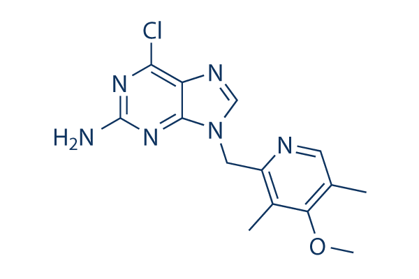
Chemical Structure
Molecular Weight: 318.76
Quality Control
Cell Culture, Treatment & Working Concentration
| Cell Lines | Assay Type | Concentration | Incubation Time | Formulation | Activity Description | PMID |
|---|---|---|---|---|---|---|
| MCF7 | Function assay | Inhibition of HSP90-mediated client protein HER2 degradation in human MCF7 cells, IC50=0.038μM | 20055425 | |||
| BT474 | Function assay | Binding affinity to Hsp90 nucleotide binding domain in human BT474 cells, IC50=0.14μM | 20608738 | |||
| MCF7 | Function assay | Inhibition of HSP90alpha in human MCF7 cells assessed as degradation of Her-2, EC50=0.038μM | 22938030 | |||
| NCI-H295 | Cytotoxicity assay | 120 mg/kg | 5 days | Cytotoxicity against human NCI-H295 cells overexpressing PGP xenografted in athymic mouse assessed as inhibition of tumor growth at 120 mg/kg, po qd for 5 days per week for 4 weeks | 22938030 | |
| Sf9 | Function assay | 3 hrs | Binding affinity to human N-terminal polyHis-tagged HSP90alpha (D9 to E236) alpha-helix conformation expressed in insect sf9 cells after 3 hrs by fluorescence polarization assay, Ki=0.002μM | 24332488 | ||
| Sf9 | Function assay | 3 hrs | Binding affinity to human N-terminal polyHis-tagged HSP90beta (D9 to E236) expressed in insect sf9 cells after 3 hrs by fluorescence polarization assay, Ki=0.004μM | 24332488 | ||
| NCI-H1299 | Function assay | 12 hrs | Reduction in oxygen consumption rate in human NCI-H1299 cells incubated for 12 hrs | 25383915 | ||
| HeLa | Function assay | 10 uM | 6 hrs | Inhibition of HSP90 in human HeLa cells assessed as induction of chk1 degradation at 10 uM after 6 hrs by Western blot method | 28816449 | |
| HeLa | Function assay | 10 uM | 6 hrs | Inhibition of HSP90 in human HeLa cells assessed as induction of Akt degradation at 10 uM after 6 hrs by Western blot method | 28816449 | |
| HeLa | Function assay | 10 uM | 6 hrs | Inhibition of HSP90 in human HeLa cells assessed as induction of HSP70 protein expression at 10 uM after 6 hrs by Western blot method | 28816449 | |
| PC3 | Function assay | 10 uM | 6 hrs | Inhibition of HSP90 in human PC3 cells assessed as induction of chk1 degradation at 10 uM after 6 hrs by Western blot method | 28816449 | |
| PC3 | Function assay | 10 uM | 6 hrs | Inhibition of HSP90 in human PC3 cells assessed as induction of Akt degradation at 10 uM after 6 hrs by Western blot method | 28816449 | |
| PC3 | Function assay | 10 uM | 6 hrs | Inhibition of HSP90 in human PC3 cells assessed as induction of HSP70 protein expression at 10 uM after 6 hrs by Western blot method | 28816449 | |
| TC32 | qHTS assay | qHTS of pediatric cancer cell lines to identify multiple opportunities for drug repurposing: Primary screen for TC32 cells | 29435139 | |||
| SJ-GBM2 | qHTS assay | qHTS of pediatric cancer cell lines to identify multiple opportunities for drug repurposing: Primary screen for SJ-GBM2 cells | 29435139 | |||
| A673 | qHTS assay | qHTS of pediatric cancer cell lines to identify multiple opportunities for drug repurposing: Primary screen for A673 cells | 29435139 | |||
| SK-N-MC | qHTS assay | qHTS of pediatric cancer cell lines to identify multiple opportunities for drug repurposing: Primary screen for SK-N-MC cells | 29435139 | |||
| NB-EBc1 | qHTS assay | qHTS of pediatric cancer cell lines to identify multiple opportunities for drug repurposing: Primary screen for NB-EBc1 cells | 29435139 | |||
| SK-N-SH | qHTS assay | qHTS of pediatric cancer cell lines to identify multiple opportunities for drug repurposing: Primary screen for SK-N-SH cells | 29435139 | |||
| NB1643 | qHTS assay | qHTS of pediatric cancer cell lines to identify multiple opportunities for drug repurposing: Primary screen for NB1643 cells | 29435139 | |||
| LAN-5 | qHTS assay | qHTS of pediatric cancer cell lines to identify multiple opportunities for drug repurposing: Primary screen for LAN-5 cells | 29435139 | |||
| Rh18 | qHTS assay | qHTS of pediatric cancer cell lines to identify multiple opportunities for drug repurposing: Primary screen for Rh18 cells | 29435139 | |||
| OHS-50 | qHTS assay | qHTS of pediatric cancer cell lines to identify multiple opportunities for drug repurposing: Primary screen for OHS-50 cells | 29435139 | |||
| Rh41 | qHTS assay | qHTS of pediatric cancer cell lines to identify multiple opportunities for drug repurposing: Primary screen for Rh41 cells | 29435139 | |||
| HCT116 | Antiproliferative assay | 48 hrs | Antiproliferative activity against human HCT116 cells after 48 hrs by sulforhodamine B assay, GI50=0.15μM | 29567459 | ||
| A549 | Antiproliferative assay | 48 hrs | Antiproliferative activity against human A549 cells after 48 hrs by sulforhodamine B assay, GI50=0.33μM | 29567459 | ||
| NCI-H1975 | Antiproliferative assay | 48 hrs | Antiproliferative activity against human NCI-H1975 cells after 48 hrs by sulforhodamine B assay, GI50=0.38μM | 29567459 | ||
| HL60 | Antiproliferative assay | 48 hrs | Antiproliferative activity against human HL60 cells after 48 hrs by sulforhodamine B assay, GI50=0.59μM | 29567459 | ||
| HL60 | Function assay | 1 uM | 24 hrs | Inhibition of HSP90 in human HL60 cells assessed as induction of HSP70 expression at 1 uM after 24 hrs by Western blot analysis | 29567459 | |
| HL60 | Function assay | 1 uM | 24 hrs | Inhibition of HSP90 in human HL60 cells assessed as downregulation of phosphorylated Akt expression at 1 uM after 24 hrs by Western blot analysis | 29567459 | |
| HL60 | Function assay | 1 uM | 24 hrs | Inhibition of HSP90 in human HL60 cells assessed as downregulation of phosphorylated STAT3 expression at 1 uM after 24 hrs by Western blot analysis | 29567459 | |
| HL60 | Function assay | 1 uM | 24 hrs | Inhibition of HDAC in human HL60 cells assessed as upregulation of acetyl-alpha-tubulin levels at 1 uM after 24 hrs by Western blot analysis | 29567459 | |
| HL60 | Function assay | 1 uM | 24 hrs | Inhibition of HDAC in human HL60 cells assessed as upregulation of acetylated histone H3 levels at 1 uM after 24 hrs by Western blot analysis | 29567459 | |
| MCF7 | Antiproliferative assay | Antiproliferative activity against human MCF7 cells, IC50=0.31μM | 31663736 | |||
| Click to View More Cell Line Experimental Data | ||||||
Chemical Information, Storage & Stability
| Molecular Weight | 318.76 | Formula | C14H15ClN6O |
Storage (From the date of receipt) | |
|---|---|---|---|---|---|
| CAS No. | 848695-25-0 | Download SDF | Storage of Stock Solutions |
|
|
| Synonyms | CNF2024 | Smiles | CC1=CN=C(C(=C1OC)C)CN2C=NC3=C2N=C(N=C3Cl)N | ||
Solubility
|
In vitro |
DMSO
: 64 mg/mL
(200.77 mM)
Ethanol : 12 mg/mL Water : Insoluble |
Molarity Calculator
|
In vivo |
|||||
In vivo Formulation Calculator (Clear solution)
Step 1: Enter information below (Recommended: An additional animal making an allowance for loss during the experiment)
Step 2: Enter the in vivo formulation (This is only the calculator, not formulation. Please contact us first if there is no in vivo formulation at the solubility Section.)
Calculation results:
Working concentration: mg/ml;
Method for preparing DMSO master liquid: mg drug pre-dissolved in μL DMSO ( Master liquid concentration mg/mL, Please contact us first if the concentration exceeds the DMSO solubility of the batch of drug. )
Method for preparing in vivo formulation: Take μL DMSO master liquid, next addμL PEG300, mix and clarify, next addμL Tween 80, mix and clarify, next add μL ddH2O, mix and clarify.
Method for preparing in vivo formulation: Take μL DMSO master liquid, next add μL Corn oil, mix and clarify.
Note: 1. Please make sure the liquid is clear before adding the next solvent.
2. Be sure to add the solvent(s) in order. You must ensure that the solution obtained, in the previous addition, is a clear solution before proceeding to add the next solvent. Physical methods such as vortex, ultrasound or hot water bath can be used to aid dissolving.
Mechanism of Action
| Targets/IC50/Ki |
HSP90
(Cell-free assay) 1.7 nM(Ki)
|
|---|---|
| In vitro |
BIIB021 binds in the ATP-binding pocket of Hsp90, interferes with Hsp90 chaperone function, and results in client protein degradation and tumour growth inhibition. This compound inhibits tumour cell (BT474, MCF-7, N87, HT29, H1650, H1299, H69 and H82) proliferation with IC50 from 0.06-0.31 μM. It induces the degradation of Hsp90 client proteins including HER-2, Akt, and Raf-1 and up-regulated expression of the heat shock proteins Hsp70 and Hsp27. This chemical inhibits Hodgkin's lymphoma cells (KM-H2, L428, L540, L540cy, L591, L1236 and DEV) with IC50 from 0.24-0.8 μM. It shows low activity in lymphocytes from healthy individuals. This compound inhibits the constitutive activity of NF-κB despite defective IκB. It induces the expression of ligands for the activating NK cell receptor NKG2D on Hodgkin's lymphoma cells resulting in an increased susceptibility to NK cell–mediated killing. This chemical enhanced the in vitro radiosensitivity of HNSCCA cell lines (UM11B and JHU12) with a corresponding reduction in the expression of key radioresponsive proteins, increased apoptotic cells and enhanced G2 arrest. It is considerably more active than 17-AAG against adrenocortical carcinoma H295R, both in vitro and in vivo. The cytotoxic activity of this compound is not influenced by loss of NQO1 or Bcl-2 overexpression, molecular lesions that do not prevent client loss but are nonetheless associated with reduced cell killing by 17-AAG. It is also active in 17-AAG resistant cell lines (NIH-H69, MES SA Dx5, NCI-ADR-RES, Nalm6 and etc.).
|
| Kinase Assay |
Hsp90 Binding Assay
|
|
For fluorescence polarization competition measurements, the FITC-geldanamycin probe (20 nM) is reduced with 2 mM TCEP at room temperature for 3 hours, after which the solution is aliquoted and stored at -80 °C until used. Recombinant human Hsp90α (0.8 nM) and reduced FITC-geldanamycin (2 nM) are incubated in a 96-well microplate at room temperature for 3 hours in the presence of assay buffer containing 20 mM HEPES (pH 7.4), 50 mM KCl, 5 mM MgCl2, 20 mM Na2MoO4, 2 mM DTT, 0.1 mg/mL BGG, and 0.1% (v/v) CHAPS. Following this preincubation, this compound in 100% DMSO is then added to final concentrations of 0.2 nM to 10 μM (final volume 100 μL, 2% DMSO). The reaction is incubated for 16 hours at room temperature and fluorescence is then measured in an Analyst plate reader, excitation = 485 nm, emission = 535 nm. High and low controls contained no this chemical or no Hsp90, respectively. The data are fit to a four-parameter curve and IC50 is generated.
|
|
| In vivo |
Oral administration of BIIB021 leads to tumour growth inhibition in many tumour xenograft models including N87, BT474, CWR22, U87, SKOV3 and Panc-1. This compound effectively inhibits growth of L540cy tumour at a dose of 120 mg/kg. It significantly enhances antitumor growth effect of radiation in JHU12 xenograft.
|
References |
|
Clinical Trial Information
(data from https://clinicaltrials.gov, updated on 2024-05-22)
| NCT Number | Recruitment | Conditions | Sponsor/Collaborators | Start Date | Phases |
|---|---|---|---|---|---|
| NCT01017198 | Completed | Advanced Solid Tumors |
Biogen |
November 2009 | Phase 1 |
| NCT01004081 | Completed | Breast Cancer |
Biogen |
November 2009 | Phase 2 |
| NCT00618735 | Completed | Advanced Solid Tumors |
Biogen |
February 2008 | Phase 1 |
| NCT00618319 | Completed | GIST |
Biogen |
February 2008 | Phase 2 |
Tech Support
Tel: +1-832-582-8158 Ext:3
If you have any other enquiries, please leave a message.
Products are for research use only. Not for human use. We do not sell to patients.
©Copyright 2013 Selleck Chemicals. All Rights Reserved.